 Neurons and Synapses
Using the new tools of microscopy, scientists started to examine many different tissues, until, in the years from 1836 to 1838 (roughly a decade after the new microscopes were available), the first cells in the nervous tissue were described by several anatomists, such as Gabriel Gustav Valentin (1810-1863) and Christian Gottfried Ehrenberg (17951876). Valentin was the first to describe the cell, nucleus and nucleolus of neurons, in 1836.
 Drawing of a neuron by Gabriel Valentin, showing the protoplasm (a), the nucleus (c), the nucleolus (d) and the axonal cone (b). |
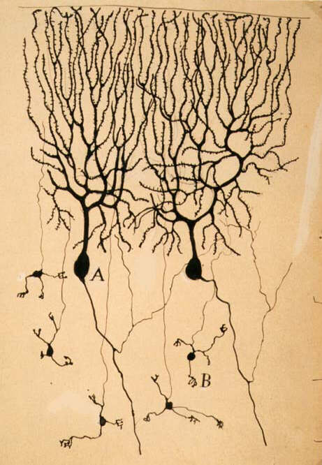
Among these pioneers, was a Czech anatomist who became a giant in the field, Jan Evangelista Purkinje (1787-1869). After waiting patiently for seven years for getting his first achromatic microscope, he started to study neurons in the cerebellum. In 1837, Purkinje described not only clusters of beautiful drop-like cells, but also subtle elongated fiber-like processes in their vicinity, which seemed to be peculiar to the nervous system. Purkinje was the first to use the microtome, potassium bichromate and Canada balsam in the preparation of histological slides for microscopy.
Another influential scientist of this period, Robert Remak (1815-1865), described
in 1836 how the nervous tissue seemed to be entirely suffused with a very fine and exceedingly complex mesh of
filamental processes, which had escaped the eyes of previous microscopists. He also described for the first time
the existence of two types of nerve processes: myelinated and unmyelinated. No one knew for sure what was the function
of these filaments, but they seemed to be important. Purkinje proposed that there should be some connection between
these processes and the nucleated cell bodies. Remak suggested in no uncertain terms that the nerve fibers could
be processes which arose from the nerve cell.
|
|
|
|
Unfortunately, the absence of techniques to stain differentially the neural cells
hindered further progress in the scientific debate about the cellular structure of the brain, until the beginning
of the century's second half. Around the 1850s, carmine red, a bright stain obtained from some insects, was used
by Alfonso Corti
(1822-1876), an Italian (who later became famous for his descriptions of the inner ear organ of hearing which bears
his name), and by Joseph von Gerlach (1820-1886), a German, to obtain the clearest images ever of neural cells
and its filaments. In addition, a much improved fixative for the nervous system, chromic acid, was discovered in
1840 by Adolph Hannover (1814-1894). Later in the century, new synthetic stains derived from anilin contributed
a lot to histological techniques of the nervous system.
|
Jan E. Purkinje in 1837. |
axons, by Otto F K. Deiters, in 1865. |
Around 1863, Otto Friedrich Karl Deiters (1834-1863), using chromic acid and carmine
red, studied extensively many nervous tissues. He also developed a microdissection technique to isolate neurons under
the microscope. He obtained remarkable clear and complete images of neuron cells, and found that there were two
different kinds of branching processes attached to the soma: one which was tree-like, full of fine, short branches,
which he called "protoplasmic extensions",
and another which was more like a long fiber, with a much smaller number of branchings, which he called "axis cylinder". These structures continued to
be studied, and received a proper name 20 years later, only: respectively "dendrites" in 1889, by Wilhelm His (1831-1904), and "axons" in 1896, by Rudolph Albert von Kölliker. The cell itself was christened only
in 1891 as "neuron", by
Heinrich Wilhelm von Waldeyer (1836-1921).
|
|
|
|
Deiters suggested cautiously that the endings of the axons of one cell could actually be joined to the dendrites of another, such as in an anastomosis, or fusion between two cells. Joseph von Gerlach, now a famous and respected histologist, proposed that the recently discovered nerve impulses ("action currents"), studied by his German colleagues Emil du Bois-Reymond and Julius Bernstein, propagated from cell to cell across these filaments, and that the brain was formed by giant nets, or reticula, with a huge number of such interconnected filaments. Von Kölliker proposed that perhaps only the dendrites were fused. We will see that von Kölliker and von Waldeyer were both very important later on in the century, for supporting the neuron doctrine, based on findings of Santiago Ramón y Cajal.
|
Drawing by Joseph von Gerlach showing neural reticulum |
Finally, 70 years since Galvani had proposed the first scientifically feasible theory about the way neural tissue works, a fascinating picture was slowly taking form. All neural functions, and even perhaps the mind itself, could be the result of the feverish transmission of electric messages throughout this huge syntitium, very much like the telegraph communication networks firmly established across the whole world. Nerve impulses, which were already known at the time to be "all-or-none" ("digital" was a term invented much later), as suggested by an American physiologist, Henry Pickering Bowditch (1840-1911), could be actually the dots and dashes of messages being transmitted. It would be the main neuroscience's task for the next centuries to understand this language as thoroughly as possible, very much like Champollion decrypted the Egyptian hyerogliphs.
|
Neurons and Synapses: The History of Its Discovery Part 2 of 6 |
 |


