

 Communication
Between Nerve Cells
By Silvia Helena Cardoso,
PhD
Introduction
All of our sensations, feelings, thoughts, motor and emotional responses, learning and memory, the actions of psychoactive drugs, the causes of mental disorders, and any other function or dysfunction of the human brain cannot be understood without the knowledge about the fascinating process of communication between nerve cells (neurons). Neurons must continuously gather information about the internal state of the organism and its external environment, evaluate this information, and coordinate activities appropriate to the situation and to the person's current needs.
How neurons process this information?
This essentially happens by means of the nerve impulse. A nerve impulse is the transmission of a coded signal from a given stimulus along the membrane of the neuron, starting in the point where it was applied. Nerve impulses can pass from one cell to another, thus creating a chain of information within a network of neurons.
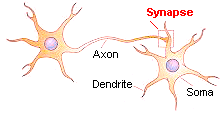
Two types of phenomena are involved in processing the nerve impulse: electrical and chemical. Electrical events propagate a signal within a neuron, and chemical processes transmit the signal from one neuron to another or to a muscle cell. The chemical process of interaction between neurons and between neurons and effector cells occur at the end of the axon, in a structure called synapse. Touching very close against the dendrite of another cell (but without material continuity between both cells), the axon releases chemical substances called neurotransmitters, which attach themselves to chemical receptors in the membrane of the following neuron and promote excitatory or inhibitory changes in its membrane.
Therefore, neurotransmitters make possible the nerve impulses of one cell influence the nerve impulses of another, thus allowing brain cells to "talk to each other", so to speak. The human body has developed a large number of these chemical messengers in order to facilitate internal communication and signal transmission within the brain. When everything is working properly, the internal communications take place without we even being aware of them.
An understanding of synaptic transmission is the key to understanding the basic operation of the nervous system at a cellular level. The whole point of the nervous system is to control and coordinate body function and enable the body to respond to, and act on, the environment. Synaptic transmission is the key process in the integrative action of the nervous system.
We have already seen the electrical process of the nerve impulse in previous article. In this issue, we are going to examine more closely how the synapse and neurotransmitters work..
Synapse: Meeting Point Between Neurons
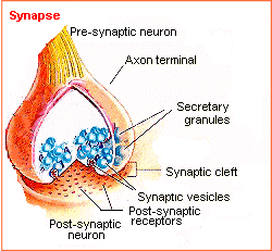
Since neurons form a network of electrical activities, they somehow have to be interconnected. When a nerve signal, or impulse, reaches the ends of its axon, it has traveled as an action potential, or a pulse of electricity. However, there is no cellular continuity between one neuron and the next; there is a gap called synapse. The membranes of the sending and receiving cells are separated from each other by the fluid-filled synaptic gap. The signal cannot leap across the gap electrically. So, special chemicals called neurotransmitters have this role. They are released by presynaptic sending membrane and seep across the gap tp receptors on the receiving neuron's postsynaptic membrane. The binding of neurotranmitters to these receptors has the effect to allowing ions (charged particles) to pass in and out of the receiving cell, as we have seen in the paper about neural conduction.
The normal direction of information flow is from the axon terminal to the target neuron; thus, the axon terminal is said to be presynaptic (carries information towards a synapse) and the target neuron is said to be postsynaptic (carries information from a synapse).
Types of synapses
The typical and most frequent type of synapse is the one in which the axon of one neuron connects to a second neuron by usually making contact with one of its dendrites or with the cell body. There are two ways in which this can happen: the electrical and the chemical synapses.
The Electrical
Synapse
 |
Most mammalian synapses are chemical, but there is a simple form of electrical synapse that allows the direct transfer of ionic current from one cell to the next. Electrical synapses occur at specialized sites called gap junctions. They form channels that allow ions to pass directly from the cytoplasm of one cell to the cytoplasm of the other. Transmission at electrical synapses is very fast, thus, an action potential in the presynaptic neuron can produce almost instantaneously, an action potential in the postsynaptic neuron. Electrical synapses in mammalian CNS, are mainly found in specialized locations where normal functions requires that the activity of neighboing neurons be highly synchronized. Although gap junctions are relatively rare between adult mammalian neurons, they are very common in a large variety of non-neural cells, including smooth cardiac muscle cells, epithelial cells, some glandular cells, glia, etc. They are also common in many invertebrates. |
The Chemical Synapse
In this type of synapse the incoming signal is transmitted when one neuron releases a neurotransmitter into the synaptic cleft which is detected by the second neuron through the activation of receptors placed opposite to the release site.
Neurotransmitters are chemicals made by neurons and used by them to transmit signals to the other neurons or non-neuronal cells (e.g., skeletal muscle; myocardium, pineal glandular cells) that they innervate.
The chemical binding
of the neurotransmitter to the receptors causes a series of physiological
changes in the second neuron which constitutes the signal. Usually the
release from the first neuron (called presynaptic) is caused by a series
of intracellular events evoked by a depolarization of its membrane, and
almost invariably when an action potential takes place.

Diagram and micrography
of a synapse of the neuromuscular junction of a fruit fly.
Photo: From Synaptic function, by Kendal Broadie, PhD, Univ. Utah. Reproduced with permission. Diagram: Silvia Helena Cardoso, PhD. Univ. Campinas, Brazil |
Synapse.
As an electrical impulse travels down the "tail" of the cell, called the
axon and arrives at its terminal, it triggers vesicles containing a neurotransmitter
to move toward the terminal membrane. The vesicles fuse with the terminal
membrane to release their contents. Once inside the synaptic cleft (the
space between the 2 neurons) the neurotransmitter can bind to receptors
(specific proteins) on the membrane of a neighboring neuron.
|

See the animation
|
What triggers the
release of a neurotransmitter?
Some mechanism must exist whereby the action potential causes the transmitter stored in synaptic vesicles to be expelled into the cleft. The action potential
stimulates the influx of Ca2+, which causes synaptic vesicles
to attach to the release sites, fuse with the plasma membrane and expel
their supply of transmitter. The transmitter diffuses to the target cell,
where it binds to a receptor protein on the external surface of the cell
membrane. After a brief period the transmitter dissociates from the receptor
and the response is terminated. In order to prevent the transmitter from
rebinding to the receptor and repeating the cycle, the transmitter is either
destroyed by degradative action of an enzime or it is taken up, usually
into the presynaptic ending. Each neuron can produce only one kind of transmitter.
|
Categories of chemical synapses
There are two
types of chemical synapses, according to the effect it causes on the postsynaptic
element:
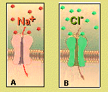
An impulse arriving in the presynaptic terminal causes the release of neurotransmitter. A.The molecules bind to transmitter-gated ion channels in the postsynaptic membrane. If Na+ enters the postsynaptic cell through the open channels, the membrane will become depolarized. B. The molecules bind to transmitter-gated ion channels in the postsynaptic membrane. If Cl- enters the possynaptic cell through the open channels, the membrane will become hyperpolarized. The resulting change in membrane potential, as recorded by a microeletrode in the cell is saw in figure below (Generation of an EPSP and IPSP). |
Excitatory
synapses:
They cause an excitatory electrical change in the postsynaptic potential (EPSP). This happens when the net effect of transmitter release is to depolarize the membrane, bringing it nearer to the electrical threshold for firing an action potential. This effect typically is mediated by the opening of membrane channels (kind of pores which traverses the cell membranes) for sodium and calcium ions. Inhibitory synapses: They cause an inhibitory postsynaptic potential (IPSP), because the net effect of transmitter release is now to hyperpolarize the membrane, making it more difficult to reach the electrical threshold potential. This type of inhibitory synapse works by opening different ion channels in the membrane: typical chloride (Cl-) or potassium (K+) channels. |
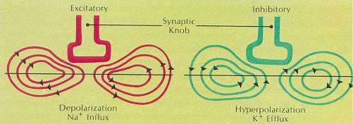

Generation of an EPSP and IPSP. |
In this figure,
the recording of the transmembrane electrical potential in function of
time (in red) shows that there is a gradual upward deflection of the trace
when an excitatory synapse is activated (EPSP). The flux of ions causes
a depolarization, i.e, the membrane becomes less polarized. Please remember
that usually the exterior face of the membrane is negative in relation
of the interior, and that the resting potential of the postsynaptic membrane
is around -70 milivolts. Any depolarization decreases this value, making
it less negative, therefore causing an upward deflection (closer to the
zero level).
The recording of the membrane potential for an inhibitory postsynaptic potential (IPSP: in green) shows a hyperpolarization, i.e., a downward deflection in the tracing, because it becomes more negative than the resting potential. A single neural cell usually has hundreds or thousands of chemical excitatory and inhibitory synapses arriving at its dendrites or cell body. EPSPs and IPSPs are summed up algebraically, so that the resulting curve (in black) may lean toward a net depolarization or hyperpolarization. If net depolarization reaches the threshold level, the postsynaptic cell fires action potentials. |
Synapses
in the central nervous system
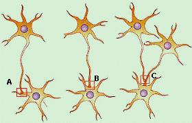 |
Different types of synapse may be distinguished by which part of the neuron is postsynaptic to the axon terminal. If the postsynaptic membrane is on a dendrite, the synapse is said to be axo-dendritic. If the postsynaptic membrane is on the cell body, the synapse is said to be axosomatic. In some cases the postsynaptic membrane is on another axon, and these synapses are called axoaxonic. In certain specialized neurons, dendrites actually form synapses with one another; these are called dendrodendritic synapses. |
Neurotransmitters: Messengers of the Brain
Chemically, neurotransmitters are relatively small and simple molecules. Different types of cells secrete different neurotransmitters. Each brain chemical works in widely spread but fairly specific brain locations and may have a different effect according to where it is activated. Some 60 neurotransmitters have been identified, and they fall mainly into one of four classes:
1) cholines; of which acetylcholine is the most important one;
2) biogenic amines: serotonin, histamine, and the catecholamines - dopamine and norepinephrine
3) amino acids - glutamate and aspartate are well known excitatory transmitters, while gamma-aminobutyric acid (GABA), glycine and taurine are inhibitory neurotransmitters.
4) neuropeptides,- these are formed by longer chains of amino acids (like a small protein molecule). Over 50 of them are known to occur in the brain, and many of them have been implied in the modulation or transmission of neural information.
Important Neurotransmitters and their Function
Dopamine
Controls arousal
levels and motor control in many parts of the brain.
When
levels are severely depleted in Parkinson's disease, patients are unable
to move voluntarily. LSD and other hallucinogenic drugs are thought to
work on the dopamine system.
Serotonin
This is the neurotransmitter
enhanced by many antidepressives, such as Prozac, and has thus become known
as the 'feel-good' neurotransmitter.It
has a profound effect on mood, anxiety and aggression.
Acetylcholine
(ACh)
Controls activity
in brain areas connected with attention, learning
and memory. People with Alzheimer's disease
typically have low levels of ACh in the cerebral cortex, and drugs that
boost its action may improve memory in such patients.
Noradrenaline
Mainly an excitatory chemical that
induces physical and mental arousal and elevated
mood. Production is centered in an area of
the brain called the locus coreuleus, which is one of several putative
candidates for the brain's 'pleasure' centre. Medical science has proven
that norepinephrine mediates the heart rate, blood pressure, the rate of
glycogen (glucose) conversion for energy, as well as and other physical
benefits.
Glutamate
The brain's major
excitatory neurotransmitter,
vital for forging
the links between neurons that are the basis of learning and long-term
memory.
Enkephalins and
Endorphins
These are opioids
that, like the drugs heroine and morphine, modulate pain, reduce stress
etc. They may
be involved in the mechanisms of physical dependence.
See Neurotransmitters: Diversity and Functions
Neurotransmitters
Overview
of Neurotransmitters and Chemical Synapses
Brain
Neurotransmitters
Neurotransmitters
- Basic Information
Molecules
of neurotransmitters
Synapse
Communication
Along & Between Neurons - Eckert & Randall - Chapter #6
Synthesis, Degradation - Neurophysiology
and Drug-Receptor Pharmacology
Vesicular
Storage - (Small Neurotransmitters )
The Author
 |
Silvia
Helena Cardoso, PhD. Psychobiologist, master and doctor in Sciences,
Founder and editor-in-chief,Brain and Mind Magazine, State University of Campinas. |