
Learning and Changes in the Brain
By Silvia Helena Cardoso, PhD and Renato M.E. Sabbatini, PhD
Until recently, neuroscientists believed that once the brain completes its development, its is unable to change, particularly in regard to its neural cells, or neurons. The dogma held that neurons cannot reproduce themselves or change significantly their connection structure to other neurons. The practical consequence for these tenets were that a) lesioned parts of the brain, such as in victims of tumors or stroke, are unable to growth again and regain part of their function; and b) experience and learning may alter the brain's functionality, but not its anatomy.
It seems that neuroscientists were wrong on both counts. Research in the last 10 years shows a strikingly different picture. In response to play, stimulation and experience, there is a growth of neural connections within the brain. Although early pioneers in biobehavioral research, such as Donald Hebb, from Canada, and Jersy Konorski, from Poland, believed that memory probably involved structural changes in the neural circuits, experimental evidence for this notion was lacking.
In experiments
carried out in rats by American neuroanatomist Dr. Marian Diamond, she
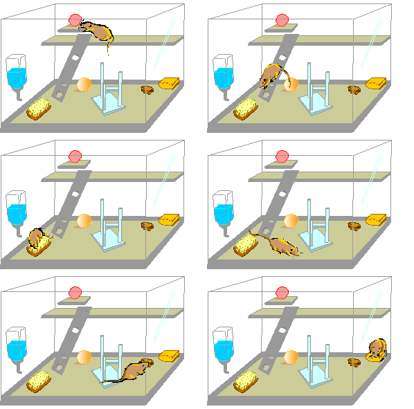
was able to show that animals that were reared in a rich environment --
a cage full of toys and features such as balls, wheels, staircases, ramps,
etc., developed a significantly thicker cerebral cortex than rats reared
in a poorer environment, without toys, or in isolation. The increased thickness
was due not only to a larger number of brain cells, but also there was
more extensive branching of their dendrites and of interconnections to
other cells.


Feature poor environment |

 Enriched environmental allowing rats to interact with toys in their cage produces anatomical changes in the cerebral cortex. |
 The
graph on the right shows how the mean number of branches observed in stellate
cells, which are found in the fourth layer of the rat's visual cortex,
increase more on animals reared on rich environments (curve labelled EC,
or enriched condition), than in animals reared together in social groups,
but with no toys (SC, or social condition group). SC rats, on the other
hand, have more branching in the stellate cells than animals reared isolated,
and without toys (IC, or isolated condition group). A larger difference
between the groups can be seen on the 4th and 5th level branches. In other
words: branches arising from the cell's body do not get increased because
of rich sensory, motor and social experience; but branches at the tip of
long processes, do. This is evidence that the stellate cells are growing
and extending new sub-branches on existing ones, and this is revolutionary
knowledge.
The parts of the stellate cells which are growing are the dendrites. It is through the dendrites that a neuron receives nervous impulses from other cells, which are conveyed to the cell's body, and thence to the axon. Therefore, a growth in dendrite branching can only mean that intercommunication processes in the cortex cells have increased and that more dendrites make them more effective in terms of regulating the activity of neural circuits.
Does this growth happens in humans, too?
It seems so, although
direct evidence, such as that observed by Diamond in rats, is not available
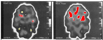
yet. However, we know that mental activation tasks are accompanied by many
changes, such as in brain metabolism (consumption of glucose by brain cells,
increase in blood flow and temperature, etc.). These changes can now be
observed directly by using computerized imaging devices, such as functional
magnetic resonance imaging (fMRI) and positron emission tomography (PET).

Cerebral Blood Flow (CBF) during a mental activation task. Normal control subject, baseline (left) and mathematical task (right). In this subject, increased perfusion during the mathematicaltask is present in both inferior frontal and left parietal areas. Source: Villanueva-Meyer et. cols. - Alasbimn Journal |
The practical consequences for the knowledge that brain cells grow and modify themselves in response to rich learning and experience are extraordinary:
Conclusions
Neural growth and regeneration in response to environmental factors no longer appear to be impossible, from what neuroscience has disclosed in experiments with animals and humans. This knowledge, coupled with the discovery of the mechanisms that make this possible will be the gateway to a fantastic future for humankind; a future where we may well be able to manipulate and influence our own mental capacities in unforeseen ways. This has been a long-standing dream of fiction and science alike, and we may be in the threshold to its realization.
The Authors

Silvia Helena Cardoso, PhD. Psychobiologist, master and doctor in Sciences, Founder and editor-in-chief, Brain and Mind Magazine, State University of Campinas. |

Renato M.E. Sabbatini, PhD. Doctor in Sciences (Neurophysiology) by the State University of São Paulo Associate Director of the Center for Biomedical Informatics of the State University of Campinas, Brazil, and president of the Editorial Board, Brain & Mind Magazine. |