
 The
Psychopath's Brain
The
Psychopath's Brain[Back to the first page]
[ A case for the frontal brain | Images of violence | Conclusions | Next ]
Why sociopaths have these characteristics ? Are their brains different from those of normal people ? Do they display pathological alterations ?
Many studies have shown in the last 20 years that murderers and ultraviolent criminals have a startling evidence of brain disease. For example, in one such study, 20 of 31 confessed or sentenced murderers had specific neurological diagnoses. Some of the inmates had more than one disorders, and no subject was normal in all spheres. Among the diagnoses were schizophrenia, depression, epilepsy, alcoholism, alcoholic dementia, mental retardation, cerebral palsy, brain injury, dissociative disorders and others. More than 64 % of them appeared to have frontal lobe abnormalities. Fifty percent had brain atrophy and 40 % had EEG abnormalities. Almost 84 % of the subjects had been victims of severe physical and/or sexual abuse. The group of murderers included gang members, rapists, robbers, serial murderers, mass murderers, one subject who killed his infant son, and another who murdered three siblings.
In another study carried out in Canada in 1994, in the most violent group of 372 males imprisoned in a maximum-security mental hospital, 20 % had focal temporal abnormalities of the EEG, and 41 % had pathologic alterations of the brain structure in the temporal lobe. The corresponding rates for the least violent group were 2.4 % and 6.7 %, respectively, thus suggesting an important role of neurological damage in the genesis of violent personalities.
According to authors Nathaniel J. Pollone and James J. Hennessy, "[Various] studies over a period of nearly 40 years... suggest a relative incidence of neuropathology among violent offenders many times in excess of that found in the general population, at ratios ranging from a high of 31:1 in the case of homicide offenders through 21:1 among `habitual aggressive' offenders to a low of 4:1 in the case of `one-time aggressives.' We propose that, though such discrepancies do not confirm neuropathology as univariately causative of criminal aggression, neither is it reasonable to believe that they are simple artifacts of chance." (35th Annual Meeting of the Academy of Criminal Justice Sciences, Albuquerque, NM, March 14, 1998)
Although this has been always a very controversial subject, many researchers think that there is now a compelling case for a substrate of brain disease in violent criminals; and this has very important consequences for many things, from the point of view of the law to the prospect of effective prevention and treatment of sociopathy.
Since sociopathic individuals have marked alterations in their relation to other human beings, it is only natural that we should first seek whether the part of their brains responsible for this has some significant abnormality.
Much of the behavior which makes possible stable and adequate social relations is controlled by the part of the brain called frontal lobe, which is located in the most anterior part of the brain hemispheres. All social primates have highly developed frontal brains, and human beings have the largest one of all. Self-control, planning, judgment, the balance of individual versus social needs, and many other essential functions underlying effective social intercourse are mediated by the frontal structures of the brain (see Dr. Silvia Cardoso's enlightening article on "The External Architecture of the Brain" in Brain & Mind Magazine's first issue, to understand what's the frontal brain).
 |
Main subdivisions of the human encephalon. The frontal areas include the frontal lobe (its anterior tip is called prefrontal area) and the motor cortex (responsible for the voluntary control of muscle movement) and sensory cortex (which receives the sensory information coming mainly from touch, vibration, pain, proprioception and temperature sensors; there are separate areas for olfaction, taste, vision and audition). Broca's area is a specialized area responsible for the motor expression of speech. |
For a long time now, neuroscientists known that lesions to this part of the brain lead to severe deficits in all these behaviors. The inordinate use of prefrontal lobotomy as a therapeutic tool by surgeons for many mental diseases in the 40s and 50s, provided researchers with enough data to implicate the frontal brain in the genesis of dissocial and antisocial personalities (see my article on the history of psychosurgery in the second issue of Brain & Mind Magazine).
 |
Illustration of trans-orbital leucotomy, a surgical operation which was widely used in the 50s to perform prefrontal lobotomies in many types of mental disease. Developed by American neurosurgeon Walter Freeman, it consisted in inserting a blade through the roof bone of one of the eye orbits using a hammer and local anesthesia. The movement of the blade severed important connections between the frontal areas and the rest of the brain. |
There are also many examples of persons who may have acquired sociopathic personalities due to pathological brain lesions, such as tumors. For example, a case report in 1992 described a patient who developed personality changes which strongly resembled an antisocial personality disorder after surgical removal of a pituitary tumor, which provoked damage to a part of the frontal lobe called the left orbitofrontal cortex. In this case, neuropsychological and personality testing revealed no specific cognitive deficits or psychopathology.
Antonio and Anna Damasio, two noted Portuguese neurologists and researchers working in the University of Iowa, have been investigating in the last decade the neurological basis of psychopathy. They have shown in 1990, for instance that individuals who had undergone damage to the ventromedial frontal cortex (and who had normal personalities before the damage) developed abnormal social conduct, leading to negative personal consequences. Among other things, they presented inadequate decision-making and planning abilities, which are known to be processed by the frontal lobe of the brain.

Computerized reconstruction |
The Damasios have also reconstituted neurologically the first known case of personality alteration due to a frontal brain lesion, observed in the 19th century. Phineas Gage, a railroad work supervisor blasted part of his own brain with a steel rod which traversed his skull when a explosive charge was set accidentally (see my article on his case in Brain & Mind Magazine, and also the website which records the 100th anniversary of the accident). He survived for many years the extensive trauma, but he became an entirely new person, abusive and aggressive, a liar and irresponsible person, incapable of insight and planning; and quite different from his former self (according to a contemporary, "Phineas was no longer Phineas"). Based on a sophisticated computerized reconstruction of the possible extent of brain damage, Gage seemed to have a lesion on the ventromedical frontal cortex, in a place very similar to those of the modern Damasio's patients. |
Why the frontal brain seems to be so important in the genesis of antisocial individuals ?
Research with animals has shown that the right orbitofrontal cortex is involved in fear conditioning. For instance, when a rat is punished with an electrical shock every time a light blinks in its cage, it develops a fear association between the stimulus and the punishment. Normal humans learn very early in life to avoid antisocial behavior because they are punished for it and because they have the brain circuits to associate fear of punishment (feeling emotion) to behavior suppression. This seems to be a key element in the development of personality. When there is no punishment, or when the person is unable to be conditioned by fear, due to a lesion in the orbitofrontal cortex, for example, or due to lowered neural activity in this area, then it develops an antisocial personality.
We have now a direct way of visualizing brain function, which has lead to a remarkable explosion in our knowledge about the inner workings of the psychopath's brain in the last two to three years:
| Functional images of the brain, such as those provided by PET (positron emission tomography) have been used to corroborate the existence of neurological deficits in the frontal lobe in sociopaths. PET shows computer reconstructed transversal sections of the brain, showing in vivid color the level of metabolic activity of neurons. This is achieved by injecting radioactivity-marked glucose molecules into the patient's blood flow and seeing how much of it is incorporated into living brain cells. The more active the cells are (when they are processing information, for example), more intense will be the image at that point (see my article "PET: A New Window Into the Brain", in the first issue of Brain & Mind Magazine, to understand better how this technique works). | 
Positron Emission Tomography (PET) scanner obtains sectional images of the living brain, using color to depict degree of activity. Crump Institute for Biological Imaging, University of California at Los Angeles. |
Using the PET technique, American medical researchers Adrian Raine and colleagues have been studying murderers, with startling results. They found that 41 murderers have a much decreased level of brain functioning in the prefrontal cortex than normal persons, indicating a deficit related to violence. In other words, even when no visible pathological alteration was present, frontal damage was apparent by a abnormal lower activity of the brain in that area. "Damage to this brain region," Raine noted, "can result in impulsivity, loss of self-control, immaturity, altered emotionality, and the inability to modify behavior, which can all in turn facilitate aggressive acts." Other abnormalities observed by the PET study of the murderers' brain included reduced neural metabolism in the superior parietal gyrus, left angular gyrus, and the corpus callosum, and abnormal asymmetries of activity in the amygdala, thalamus, and medial temporal lobe. It is probable that these effects are related to violence and criminality; because some of these structures are part of the so-called limbic brain, which processes emotions and emotional behavior (please see "Limbic System: The Center of Emotions" in Brain & Mind Magazine)
One interesting aspect of Dr. Raine's research is that he correlated the PET brain images to the murderer''s personal history, in order to ascertain whether they were subjected to trauma, physical or sexual abuse, neglect, poverty, when they were children (a deprived environment for the development of personality). Of the murderers, 12 had suffered significant abuse or deprivation. It was discovered that murderer's coming from non-deprived households had much larger deficits in the orbitofrontal area of the brain (14 % on the average) than normal people and murderer's coming from deprived environments (see figure below).
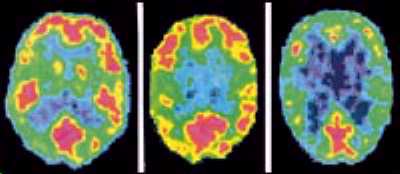 |
| PET images of the brain of a normal person (left), a murderer with deprived background (middle) and a murderer with non-deprived background (right). Areas in red and yellow show a higher metabolic activity, and in black and blue of lower metabolic activity. The brain of a sociopath (right) has a very low activity in many areas, but which is strikingly absent in the frontal area (upper part of the images). Images by Adrian Raine, University of Southern California, Los Angeles, USA. |
The initial controlled studies carried out by Raine and colleagues have been confirmed by a series of PET-based investigations with sociopathic individuals and violent criminals. In a study in 1994, 17 patients with diagnoses of personality disorder were subjected to PET scams. The researchers proved that there was a strong inverse correlation between a life history of aggressive impulse difficulties and regional metabolism in the frontal cortex. Six of these patients were antisocial, the rest had several personality disorders (borderline, dependent and narcissistic). PET was used again in 1995 to evaluate brain glucose metabolism in eight normal subjects and eight psychiatric patients with a history of repetitive violent behavior. The authors observed that "seven of the patients showed widespread areas of low brain metabolism, particularly in medial temporal and prefrontal cortices than did normal comparison subjects. These regions have been implicated as substrates for aggression and impulsivity, and their dysfunction may have contributed to the patients' violent behavior". More recently (1997), PET brain imaging technology found that psychopaths differed from nonpsychopaths in the pattern of relative cerebral blood flow during processing of emotional words. Acquired personality changes due to brain injury are also accompanied by a decrease in the neural activity in the frontal area (see figure below).
 |
A PET image of decrease in neural activity in the frontal area (upper part of the images) of the brain of a patient who sustained closed head injury (1A), and developed personality changes. Figure 1B shows a normal brain in the same area. Brain Imaging Center, University of California at Irvine |
Indirect evidence of the role of prefrontal cortex in psychopathic behavior is coming from other experiments as well. In Canada, a team headed by Dominique LaPierre compared 30 psychopaths to 30 non-psychopathic criminals, using tests that evaluate the function of two parts of the prefrontal cortex: the orbitofrontal and the frontal ventromedial areas. The results showed that "the psychopaths were significantly impaired on all the orbitofrontal- ventromedial tasks", but not in the function of other areas of the frontal cortex. The similarities between psychopaths and patients with prefrontal cortex damage surfaced in several areas of the study. "Both the psychopath and the orbitofrontal or ventromedial frontal patient show an exaggerated preoccupation with sexual matters, acting in a promiscuous and impersonal maladaptive way," observed the researchers. "Both are remarkable for their lack of social and ethical judgment. Both neglect long-term consequences of their actions, choosing immediate gratification over careful planning."
In summary, although many of these results must be regarded in caution, they all converge to an important discovery that the brains of violent criminals and sociopaths are indeed altered in subtle ways which can now be unveiled by new and sophisticated techniques. One important consideration is that human behavior is extremely complicated and results from an interplay of many subtle social, biological and psychological factors. "There are a lot of factors involved in crime. Brain function is just one of those," says Prof. Adrian Raine. "But by understanding the brain function, we will be in a much better position to understand the complete causes of violent behavior".
Another drawback of retrospective studies (that is, done after the disorder appeared in the studied individuals), is that it is sometimes difficult to separate cause from consequence. In other words, is the observed brain deficits the cause of the psychological abnormality or the result of it ?). In addition, the results are still preliminary and do not lend credibility to the use of these neuroimaging and function assesment methods to "diagnose" individuals at risk of sociopathy; so they should not be used for clinical or forensic purposes at the present stage.
So, there is a reasonable body of coherent evidence that sociopaths have a dysfunction of the frontal brain. Why and when this dysfunction appears is totally unknown, thus far.
![]() Emotionally Insensitive
Emotionally Insensitive
Copyright 1998 Universidade
Estadual de Campinas
Brain & Mind Magazine, Campinas,
Brazil
Center for Biomedical Informatics