
Renato M.E. Sabbatini, PhD
In the first decades of the 19th century, the budding science of neurophysiology was breaking fascinating ground. It was the first time in the history of science that a close cooperation between physics and biology was necessary, not only because biology was looking upon the phenomena of neural function under the light of the recent knowledge provided by physics, but also because they were using the same measuring instruments, devices and apparatuses.
The applied knowledge amassed by experimental physicists in the fields of electricity, optics and mechanics was starting to make a great contribution to physiology. Many times, however, the physiologists who were carrying out pioneering experiments were forced to invent or to adapt existing instruments in order to fit the demanding requirements of working with living tissue, capable of generating extremely feeble currents and voltages, never before studied by physicists.
In the first place, physiologists needed stable and reliable sources of electrical current, in order to stimulate nerves and muscles. When Luigi Galvani started his path-breaking experiments, only a few techniques were available: Leyden jars, electrostatic generators and natural electricity (namely, lightnings !).

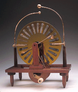
 The
electrostatic generator was made of a vertical or horizontal glass disc which could be rotated quickly using a
hand crank and belt contraption. A metallic brush, in contact with the disc, built and collected electric charges
created by attrition. Two metal balls connected to the poles were then used to transfer this charge to a Leyden
jar.
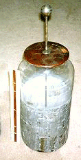
The Leyden jar was also made of glass, coated with tin foil on the inside and
a metal pin inserted through the isolated lid. It was an electric condenser, used to store electric charges as
well as to deliver them to the subject being stimulated. Unfortunately, the amount of current was not controllable
or quantifiable, thus Leyden jars were unreliable for performing repeatable experiments.
 The
definitive invention came directly from Galvani's scientific
dispute with his colleage, Alessandro Volta. Volta interpreted correctly
that Galvani's frogs would twitch their muscles when hanging from a copper hook attached to a iron railing because
the bimetal junction acted as a electricity generation device. Volta assembled then a device, the pile, with a
series of alternating silver and zinc disks, separated by pasteboard disks saturated in salt water. A current was
produced when the uppermost disk of silver was connected by wire to the bottom disk of zinc.
 The
voltaic cell battery started a sweeping technological revolution in the years to come, not only in physics but
in physiology as well. By carefully controlling the area and material of its metal parts, chemical substances and
number of elements, precise and known voltages could be delivered to the tissues being stimulated. Claude Bernard, the famous French physiologist,
fabricated very ingenuous "electrical tweezers", which he used to touch and hold delicate nerves, stimulating
them at the same time.

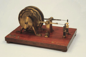
 Later
on, physiologists had to invent mechanical switches, in order to deliver pulses of electric current to nerves and
muscles at precise instants and with a very short duration. They were usually made of pools of mercury, spiked
metal rods and spring-loaded buttons. When a repeated stimulation was required by the experiment, a rotating wheel
would drive the switch at precise repetition rates, such as the Mateucci's commutator depicted here.
Electrical currents and potentials generated by biological tissues are very small, in the order of micro- and milivolts. Therefore, it is truly amazing that the primitive apparatuses used by physiologists in the 19th century could record such small variations. In one occasion, a physiologist was able to discern a mere 10 mV positive variation in the "nerve action current", which even today is difficult to see using modern electronic amplifiers and oscilloscopes.
 The
first physical device used to detect bioelectricity was the foil electrometer. This was a isolated glass container,
with two very thin gold foils attached at an angle to a connecting rod. A small electric potential applied to this
rod charged the foils with the same amount of static electricity, thus making them to repel each other. As a result,
the angle between them increased. However, the foil electrometer would not allow precise measurements.

 A better measuring instrument after
1840 was the galvanometer (its name, of course, is a hommage to Galvani). It used the electromagnetic principle.
It consisted of a tightly wound coil of isolated copper wire around a magnet glued to a needle. Passing an electric
current through the coil provoked a electromagnetic field which interacted with the field of the magnet, causing
it to turn around an axis. The amount of deviation recorded by the needle over a printed scale allowed for the
precise measurement of electric current. The larger the coil and the recording magnet, the more sensitive was the
instrument. The Swiss-German scientist Du Bois-Reymond is credited with the invention of one of the best galvanometers
used for many decades in neurophysiological experiments. His sensitive device required almost 24,000 turns of wire
in its coil, with more than 5 km in lenght!

 Later in the century, time recordings
of physiological phenomena became possible with the drum kymograph. The galvanometer's needle was put into contact
with a loop of paper coated with a thin layer of smoke black and stretched over a metal drum. A precise mechanical
clockwork allows the drum to be rotated at a calibrated speed, and the movements of the galvanometer's pen scratch
out the smoke black, leaving a record of amplitude in function of time. Mechanical phenomena, such as the contraction
of a muscle, respiratory movements and blood pressure could also be recorded along time, by rigging ingenuous contraptions
made of levers, axes, membranes, springs and strings. The time axis was calibrated and measured by using electromagnetic
tuning forks attached to inscribing pens.
 Until
the beginning of the 20th century, these were the basic tools of the electrophysiologist. The galvanometer was
perfected in 1901 by Dutch physiologist Willem Einthoven, in order to permit the measurement
and recording of extremely small variation in electric potential. It worked in the following way: silver coated
conducting string was anchored midway between two magnetic poles. The end of the string was connected to a liquid
filled electrode jar. As electrical changes occurred in the body, the string oscillated from side to side between
the magnets. . A beam of light was shone onto the silver coating, and any small angle of deflection was magnified
many times and projected onto a scale. A photorecording galvanometer consisted of a strip of photographic film
which would be displaced past the light beam, providing a time recording of the electrical phenomenon. The first
practical electrocardiograph machine was developed by Einthoven, using such a contraption.
 Click on the image to see the legends
The definitive electrophysiologist's weapon where the cathode ray tube (CRT) oscillograph
and the vacuum-tube amplifier, both invented at the turn of the century. Max Dieckmann demonstrated the principles
of CRT in Germany in 1906, and A. A. Campbell Swinton developed the concept into a practical electronic system
to record electrical variations onto a phosphor-coated screen. A heated cathode wire generates electrons, which
are directed to the screen by two pairs of charged plates: one horizontal and one vertical. The amount of deflection
of the electron beam in the vertical axis is proportional to the electrical charge applied to these plates. Uniformly
increasing voltage applied to vertical pair causes a vertical line to be drawn on the tube. American physiologists
Erlanger and Gasser made use of this new marvels of technology, electronics, at great lenght to carry out a number
of elegant and pathbreaking studies on the nerve conduction. But this is will be the subject of another article
in this series.

 The close study of the
minute structure of nerves and neurons was made possible by the invention of the microscope. Primitive microscopes
were developed by Anton van Leeuwenhoek, in the Netherlands, and Robert Hooke, in England, and both studied the
microscopic structures of nerves. However, only with the invention of the compound microscope (using several lenses
to achieve higher power) in the 18th century that anatomists and physiologists could explore in greater detail
the nervous system and discover things like the axon, the dendrites, the neuromuscular plaque, etc.
 New
staining methods, such as those developed by Santiago Ramon y Cajal and Camilo Golgi, were able to show in exquisite
detail the fine structure of the nervous tissue and its unique organization.
In this way, the modern neuron doctrine was born.
Picture credits: Museum of Physics of the University of Pavia, The Gemmary Collection, The University of Toronto Museum of Psychological Instruments.
The Field Advances![]()
 To
Know More
The Discovery of Bioelectricity
Renato M.E. Sabbatini
Brain & Mind Magazine 2(6), June/September
1998
Copyright (c) 1998 The State University of Campinas,
Brazil
Center for Biomedical Informatics