Connections of the Basal Ganglia
The basal ganglia form a collaborative system of connections between the cerebral cortex and the thalamus.
It should be noted that terms such as "strio-, pallido-, nigro-, subthalamo-" and "-striate, -pallidal, -nigral, and -subthalamic" are frequently used as prefixes and suffixes to describe pathways arising from and terminating in the respective nuclei. For example, "striatoonigral" fibers arise from the striatum (caudate + putamen) and terminate in the substantia nigra whereas "nigrostriate" fibers persue the opposite course.

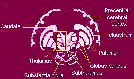
schematic representation summarizing
the connections of the
basal ganglia (see the text below)

Afferent connections |
 Corticostriatal fibers:
Thalamostriatal fibers- originate in the centromedian nucleus and in various intralaminar and midline nuclei. Nigrostriatal fibers- arise from both the pars reticularis of the substantia nigra and connect with the caudate nucleus, putamen and globus pallidus, as well as with the thalamus, frontal cortex, amygdala, superior colliculi and olfactory tubercle. The substantia nigra receives multiple input from the globus pallidus, cerebral cortex, dorsal raphe nucleus, amygdaloid body, subthalamus septal area and stria terminalis. Neurons in the substantia nigra pars reticulata (SNr) project principally to the thalamus (ventral anterior, ventral lateral, and dorsomedial nuclei) but also to brain stem nuclei (superior colliculus, pedunculopontine nucleus) and use GABA as the neurotransmittter. The SNr also receives striatal input that uses GABA (and substance P) as transmitters and is inhibitory. Amygdalostriatal fibers - projections from the basolateral amygdala to ventromedial region of the caudate nucleus and to putamen suggests that the striatum may be divided into limbic and non-limbic. |
Efferents Connections |
Caudate projects
to the: Putamen projects
to the : Globus pallidus projects
to the: |
 Efferent
fibers from corpus striatum leave via the globus pallidus. Some fibers
pass through the internal capsule
(broad band of white substance separating the lenticular nucleus from the
medial caudate nucleus and thalamus) and on reaching the medial side form
a bundle, the fasciculus lenticularis.
Other fibers sweep the medial border of the internal capsule to form a
loop, the ansa lenticularis
(from the Greek word loop) which "loops"
around the internal capsule. Both of these
sets of fibers give off some terminals to the subthalamic nucleus; others
continue upwards to the thalamus via the fasciculus thalamicus.
Efferent
fibers from corpus striatum leave via the globus pallidus. Some fibers
pass through the internal capsule
(broad band of white substance separating the lenticular nucleus from the
medial caudate nucleus and thalamus) and on reaching the medial side form
a bundle, the fasciculus lenticularis.
Other fibers sweep the medial border of the internal capsule to form a
loop, the ansa lenticularis
(from the Greek word loop) which "loops"
around the internal capsule. Both of these
sets of fibers give off some terminals to the subthalamic nucleus; others
continue upwards to the thalamus via the fasciculus thalamicus.
A major efferent route is from the globus pallidus via the ansa lenticularis to the cerebral nuclei and brain stem nuclei. The globus pallidus and the lateral nuclear groups of the thalamus appear to be focal structures upon which many pathaways concerned with motor function converge. These nuclei exert important regulating and controlling influences on motor integration, in addiction to relaying afferent systems to the cerebral cortex. The extrapiramidal system is a functional unit dependent upon an intact lateral corticospinal or pyramidal system.
In man, therapeutic production of lesions of the globus pallidus may reduce tremor and rigidity of patients with parkinsonism. (Chusid and McDonald,1967). In addiction to Parkinson's disease, pathology in the basal ganglia may lead to one of a number of motor dysfunctions known as dystonias (abnormal changes in muscular tone) and dyskinesias (uncontrolled movements).
Author: Dr. Silvia Helena Cardoso, PhD. Psychobiologist, master and doctor in Sciences. Post doctoral at University of California, Los Angeles.
Center for Biomedical Informatics
State University of Campinas, Brazil
Copyright 1997 State University of Campinas