
Neurobiology of Dreams: Neural Mechanisms
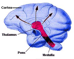
Fig.1. Monkey brain depicting
the brain stem reticular formation. In pink:
Part
|
In a higher animals and human
beings, the switching of states of awake and sleep happens due to existence
of special nervous structures.
Rem sleep, the "dream sleep", is quite different from non-REM sleep. The cortex in this state is as active during REM sleep as it is during waking. The cortex is not necessary for the production of REM, but it certainly is required for dream's elaboration. The brain activity during REM, begins in the pons, a structure in the brainstem and neighboring midbrain regions. The pons sends signals to the thalamus and to the cerebral cortex, which is responsible for most thought processes. It also sends signals to turn off motor neurons in the spinal cord, causing a temporary paralysis that prevents movement. |
Some researchers have used positron emission tomography (PET) to study the brain state associated with REM sleep in humans (Maquet et al, 1996 - da Internet). The results show that regional cerebral blood flow is positively correlated with REM sleep in:
Biochemical Mechanisms of REM sleep
Scientists found that sleep phases are closely related to the activity of certain groups of nerve cells releasing brain chemicals that relay information from one neuron to another. Research on these specialized cell groups is helping scientists to devise specific drug treatments for sleep disorders.
The brain has several colletions of neurons, each using a particular neurotransmitter and making difuse connections. These cells perform regulatory functions influencing a large number of postsynaptic neurons in spinal cord, thalamus, cerebral cortex, and so on, so they become more or less exitable. Different systems (such as noradrenergic, serotonergic and cholinergic) appear to be essential for aspects of metabolic state, motivation, motor control, memory, etc.
In his article, Hobson (bear,
1996) wrote that the most surprising observation that he and his coworkers
made was that both the noradrenergic locus
coeruleus and the serotonergic
raphe neurons "turn
off" in
REM sleep (table 1). This finding was a surprise
because every one had predicted the opposite. For him, It was clear that
these two aminergic systems supported waking and not sleep, as had been
theorized. REM sleep could be triggered by cholinergic stimulation of the
reticular formation. It is probably the action of ACh during REM sleep
that causes the thalamus and the cortex to behave so much like they do
in the wake state.
| Noradrenergic
(Locus coeruleus) |
Serotonergic
(Raphe nuclei) |
Cholinergic (cortex, septal nucleus, striatum) |
|
|
|
|
|
|
|
|
|
|
|
Table 1. Differential activity of specific neurochemical subsystems of the brain stem in waking and REM sleep. The firing rates of the two major systems of the upper brain stem, the locus coeruleus and the Raphe nuclei , "turn off" the REM sleep, that is, they decrease to almost nothing with the onset of REM. However, there is a current sharp increase in the firing rates of ACh-containing neurons in the pons, and some evidence suggests that cholinergic neurons induce REM sleep. It is probably the action of ACh during REM sleep that causes the thalamus and cortex to behave so much like they do in waking state (Bear, 474). REM periods are terminated when norepinephrine and ACh-containing neurons begin firing again.
Why Don't We Act Out During REM Sleep? (next page)

Translation: SIlvia Helena Cardoso
Center for Biomedical Informatics
State University of Campinas, Brazil
Silvia
Helena Cardoso, PhD
Contact
Copyright 1997 State University of Campinas