![[Brain & Mind Header and Navigation Bar]](../../arttec_i.jpg)
![[Brain & Mind Header and Navigation Bar]](../../arttec_i.jpg)
In the sixties, scientists working with radioactive substances, discovered a new use for them. A kind of imaging device, called a gamma camera, was invented, in order to detect the radiation emitted by breaking radioactive atoms. For example, if the physician wants to know whether the thyroid gland is working properly, he injects into the patient's bloodstream a substance attached to radioactive iodine. Iodine is easily captured and used by the thyroid gland, since its hormones have this element in its composition. The gamma camera detects every time a radioactive iodine atom emits a burst of energy (the gamma radiation), and shows it on a flat map of the patient's thyroid. After a time, the image fills with dots, and their density is higher in the regions where the thyroid cell metabolism is higher (that is, where the cells are working more actively). In this way, we achieve a functional map. A person who has a gland working improperly (too much or too few activity) will show a different map, allowing physicians to diagnose the cause and type of disease.
The PET works according to a similar principle. However, it has a highly sophisticated technique for building a image of the patient's body.

A cyclotron for the synthesis of radiopharmaceuticals
|
 The first step when making PET images of the brain, is to inject the patient
with a dose of a radiopharmaceutical. This is
a substance that can be absorbed by certain cells in the brain, concentrating
it there. For example, FDG (fluorodeoxyglucose) is a normal molecule
of glucose, the basic energy fuel of cells, attached artificially to an
atom of radioactive fluor. This element must be produced by a complex apparatus
called a cyclotron, which has also an unit to
synthetize the FDG molecule.
The cells in the brain which are more active in a given period of time after the injection, will absorb more FDG, because they have a higher metabolism and need more energy. The fluor atom in the FDG molecule suffers a radioactive decay, emitting a positron (that's a kind of electron, with a positive electrical charge, so it's anti-matter). When a positron colides with an electron, a matter-anti-matter anhilation occurs, liberating a burst of energy, in the form of two beams of gamma rays, in opposite directions. This will then be shown as an image by the PET scanner. That's why it's calle Positron Emission Tomography. |

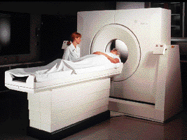
The PET scanner |
After injecting the radiopharmaceutical, the patient is
placed on a special moveable bed. which slides by remote control into the
circular opening of the scanner (called gantry). Placed around this
opening, and inside the gantry, there are several rings of radiation
detectors. Each crystal detector emits a brief pulse of light every time it
is struck with a gamma ray coming from the radioisotope within the patient's
body. The pulse of light is amplified (increased in intensity), by a photomultiplier,
and the information is sent to the computer which controls the apparatus.
The whole process is called scintigraphy (from scintillation, which
is the pulse of light).
|
![[PET color vs gray level imaging]](petcolor.gif)
A PET slice of the brain
Slice orientation |
The computer working with the scintigraphy data, reconstructs
the exact places inside the brain where each pulse of radiation came from.
It also counts the number of pulses per second coming from each point of
the image. That's because the brain structures which have higher concentrations
of the injected radiopharmaceutical emit a higher amount of radiation,
meaning that they are more active in terms of cell metabolism or blood
circulation.
The number of radiation pulses counted by the computer during a fixed interval of time is displayed in its video screen as a dot, with its intensity shown in shades of gray (gray levels). Black means no activity (zero counts), and pure white the highest count level. The same image can be displayed in false color, which is able to show in a better contrast the "hot" regions. The false color scale converts each level of gray into a shade of color, like in a rainbow, being red the highest activity count, then coming yellow, then green, and so forth. Blue, violet and black represent the lowest levels of activity. |
![[Dopa imaging of the brain with PET]](dopa.gif)
The activity of DOPA receptors in the brain |
One of the tricks used to show specific activities in the brain is to use radiopharmaceuticals which attach themselves chemically to certain neurones. In this example, a radiopharmaceutical which has a strong affinity for cells containg the DOPA transmitter, concentrates in an area of the brain called basal nuclei, which are responsible for the control of movement. These cells are damaged in Parkinson's disease, so that they have a lower concentration of DOPA. PET is an excellet method to quantify the brain function in persons with this disease. |
![[3D PET images of the brain]](pet3d.gif)
A 3D reconstruction of PET images |
Taking several adjacent slices at a time, a special computer
program can be used to make a three-dimensional reconstruction of the brain,
following any orientation or perspective desired by the physician. These
are useful for understanding better the overall image of the brain, and
what functional problems are present.![[Sequence of rotating 3D PET brain images]](sequenc2.gif)
Click on the image to see a full sequence Putting these different views as the frames of a movie, the PET computer can show it as an animated rotation. |
Image credits: The Crumb Institute of Biological Imaging, Department of Pharmacology, University of California at Los Angeles. CTI The Niels Bohr Institute
From: The PET Scan: A New Window Into the
Brain
By: Renato M.E. Sabbatini,
PhD
In: Brain & Mind Magazine, March 1997.